
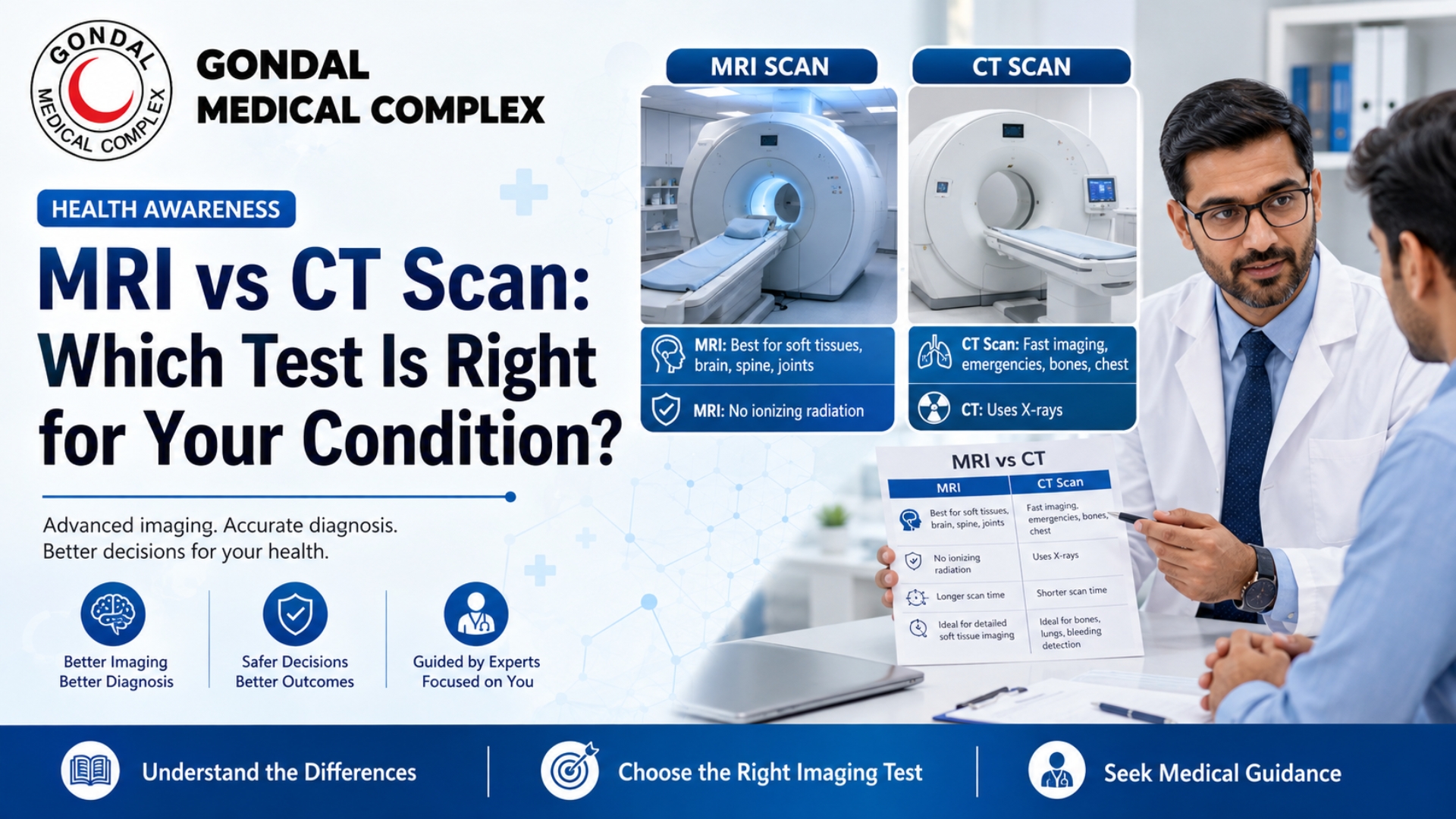
MRI vs CT Scan: Which Test Is Right for Your Condition?
When a doctor recommends diagnostic imaging, patients often wonder whether they need an MRI or a CT scan. Both tests create detailed images of structures inside the body, but they use different technologies and are suited to different medical situations.
The main MRI vs CT scan difference is that a CT scan uses X-rays to create cross-sectional images, while an MRI uses a strong magnetic field, radio waves and computer processing. CT is generally faster and is commonly used in emergencies, trauma, suspected internal bleeding and lung conditions. MRI usually provides more detailed images of the brain, spinal cord, nerves, muscles, ligaments, tendons and other soft tissues without using ionizing radiation.
Neither test is universally better. The right examination depends on the body part being assessed, the suspected condition, how urgently results are required, whether contrast is needed and whether the patient has pregnancy, kidney disease, implanted devices or difficulty remaining still.
Medical disclaimer: This article provides general educational information. The most suitable imaging test must be selected by a qualified doctor or radiologist after reviewing the patient’s symptoms and medical history.
What Is an MRI Scan?
MRI stands for Magnetic Resonance Imaging. It uses a powerful magnetic field, radiofrequency energy and computer processing to create detailed cross-sectional images of organs and tissues.
Unlike CT and conventional X-rays, MRI does not use ionizing radiation. It is particularly effective at showing differences between healthy and abnormal soft tissues. Doctors commonly use MRI to examine the brain, spine, joints, muscles, nerves, blood vessels, pelvic organs and selected abdominal structures.
During an MRI, the patient lies on a table that moves inside the scanner. The machine produces loud tapping and knocking sounds, so hearing protection is normally provided. The patient must remain still because movement can blur the images.
Common Reasons for an MRI
MRI may be selected to investigate:
- Brain or spinal-cord disorders
- Disc problems and nerve compression
- Ligament, tendon and cartilage injuries
- Joint injuries involving the knee or shoulder
- Brain tumours and other soft-tissue masses
- Multiple sclerosis and selected neurological conditions
- Spinal infections or tumours
- Pelvic and reproductive-organ conditions
- Certain blood-vessel abnormalities
- Follow-up of selected cancers
MRI is especially useful for evaluating the spinal cord, nerves, discs, ligaments, tendons, muscles and cartilage because these tissues may not appear as clearly on CT or conventional X-rays.
What Is a CT Scan?
CT stands for Computed Tomography and is also called a CAT scan. It uses rotating X-ray equipment and computer processing to create cross-sectional images of the body.
CT can show bones, internal organs, soft tissues, lungs and blood vessels in considerable detail. Images can also be reformatted into different planes or three-dimensional views.
CT examinations are generally quick and less sensitive to movement than MRI. This makes CT particularly valuable in emergency departments, where doctors may need to identify internal bleeding, serious injury, stroke or other urgent conditions without delay.
Common Reasons for a CT Scan
CT may be used to investigate:
- Head injuries
- Bleeding inside the brain
- Stroke
- Chest, abdominal or pelvic trauma
- Complicated bone fractures
- Lung infections and lung nodules
- Kidney stones
- Internal bleeding
- Blood clots in the lungs
- Certain tumours
- Blood-vessel abnormalities
- Injuries involving multiple organs
CT is particularly useful when doctors need to examine bone, lungs, blood vessels and internal organs quickly.
MRI vs CT Scan: Main Differences
| Feature | MRI | CT scan |
|---|---|---|
| Technology | Strong magnetic field and radio waves | X-rays and computer processing |
| Ionizing radiation | No | Yes |
| Speed | Usually takes longer | Usually faster |
| Soft-tissue detail | Generally excellent | Good, but often less detailed than MRI |
| Bone imaging | Useful in selected cases | Generally excellent for complex bone detail |
| Lung imaging | Limited for many routine lung conditions | Commonly preferred |
| Brain imaging | Excellent for detailed brain tissue assessment | Often preferred first in emergencies |
| Spine and nerves | Usually provides greater detail | Useful for trauma and bone structure |
| Movement sensitivity | More sensitive to movement | Less sensitive to movement |
| Metal and implants | Requires careful safety screening | Usually less restricted |
| Claustrophobia | Can be challenging | Generally quicker and more open |
| Typical contrast | Gadolinium-based contrast | Iodine-based contrast |
MRI generally provides clearer soft-tissue contrast, while CT is fast and can image bone, organs, soft tissues, lungs and blood vessels during one examination.
Which Is Better for the Brain: MRI or CT?
Both tests can examine the brain, but the preferred test depends on the suspected condition and how urgently imaging is needed.
CT for Head Injury and Emergency Bleeding
A head CT can quickly identify:
- Bleeding inside or around the brain
- Skull fractures
- Serious head injuries
- Selected signs of stroke
- Swelling or pressure-related changes
In emergency cases, CT can provide information quickly enough to guide urgent treatment. It is less sensitive to movement than MRI, which is helpful for severely injured, confused or restless patients.
MRI for Detailed Brain Assessment
Brain MRI provides detailed images of brain tissue and may help doctors evaluate:
- Brain tumours
- Multiple sclerosis
- Certain small or early strokes
- Infections and inflammation
- Pituitary abnormalities
- Seizure-related structural problems
- Cranial nerves
- Long-term neurological symptoms
MRI is often selected when detailed evaluation of brain tissue is more important than obtaining the fastest possible scan.
MRI or CT for a Suspected Stroke?
A stroke is a medical emergency. A head CT or MRI may be used to determine whether symptoms are caused by a blood clot or bleeding in the brain.
CT is frequently used as an initial emergency test because it can rapidly identify bleeding. CT angiography and CT perfusion may also be used to assess blood vessels and blood flow. MRI can provide detailed information about brain damage and may detect certain areas of early ischaemic injury. The selection depends on the patient’s condition, hospital protocol, equipment availability and the type of information needed immediately.
Sudden facial weakness, arm weakness, speech difficulty, confusion, vision loss, severe unexplained headache or loss of balance requires emergency assessment. Imaging should not be delayed while trying to decide independently between MRI and CT.
Which Is Better for the Spine?
MRI is generally preferred when doctors need to examine:
- Spinal cord
- Nerve roots
- Herniated discs
- Ligaments
- Disc infection
- Spinal tumours
- Nerve compression
- Soft tissues surrounding the spine
MRI provides detailed images of the spinal cord and nerves and can detect abnormalities that may be hidden by bone on other imaging tests.
CT may be more suitable when doctors need to assess:
- Complex spinal fractures
- Bone alignment after trauma
- Detailed vertebral bone structure
- A patient who cannot safely undergo MRI
- Selected postoperative changes
- Urgent trauma when speed is important
In some injuries, CT is performed first to examine the bones, followed by MRI when spinal-cord, ligament or nerve injury is suspected.
Which Is Better for Bones and Fractures?
Plain X-rays are usually the first imaging test for many suspected fractures. CT or MRI may be ordered when the X-ray is inconclusive or the injury is complicated.
CT for Complex Bone Detail
CT can provide detailed images and three-dimensional reconstructions of complicated fractures. It is particularly useful for injuries involving the spine, pelvis, face or joints where doctors need to understand the exact position of bone fragments.
MRI for Hidden Fractures and Soft-Tissue Injury
MRI may detect subtle fractures that do not appear on an initial X-ray. It can also show damage to surrounding ligaments, tendons, muscles, cartilage and bone marrow.
For example, when a hip fracture is strongly suspected but is not visible on an X-ray, MRI is generally considered the next best test. CT may be used when MRI is unavailable, unsuitable or difficult for the patient.
MRI or CT for Knee and Shoulder Injuries?
MRI is generally more useful for injuries involving:
- Ligaments
- Meniscus
- Tendons
- Muscles
- Cartilage
- Labrum
- Rotator cuff
- Bone marrow swelling
MRI can show many tendon, ligament, muscle and cartilage abnormalities that are less visible on CT or X-rays.
CT may be selected when the primary concern is a complex fracture, bone alignment or detailed assessment of the joint’s bony structure.
Which Is Better for the Lungs and Chest?
CT is commonly preferred for evaluating the lungs because it can provide detailed images of lung tissue and detect small abnormalities.
A chest CT may help evaluate:
- Lung nodules
- Pneumonia complications
- Tuberculosis-related changes
- Interstitial lung disease
- Bronchiectasis
- Chest tumours
- Rib or chest trauma
- Blood vessels in the chest
- Suspected pulmonary embolism when CT angiography is appropriate
Chest CT can detect very small lung nodules and is used to examine unexplained cough, shortness of breath, chest pain and abnormalities found on chest X-rays.
MRI is not usually the first choice for routine lung imaging because air-filled lungs are more difficult to examine with standard MRI techniques. However, MRI may provide useful information about the heart, blood vessels, chest wall, mediastinum and selected tumours or soft-tissue abnormalities.
MRI or CT for Abdominal Pain?
Either MRI or CT may be selected depending on the suspected cause of abdominal pain.
CT is frequently used for urgent abdominal conditions because it is fast and can examine organs, bowel, blood vessels, bones and surrounding tissues. It may help identify internal bleeding, bowel obstruction, kidney stones, appendicitis complications, abdominal trauma or selected infections.
MRI may be used when doctors need more detailed soft-tissue information or wish to avoid ionizing radiation. It can be particularly useful for selected liver, pancreas, bile-duct, pelvic and reproductive-organ conditions.
Ultrasound may also be the first test for gallstones, pregnancy-related concerns and several pelvic or abdominal conditions. The correct examination depends on the location of pain, age, pregnancy status, laboratory findings and clinical examination.
MRI or CT for Cancer?
Both MRI and CT play important roles in cancer diagnosis, staging, treatment planning and follow-up.
CT may be used to:
- Examine the chest, abdomen and pelvis
- Identify enlarged lymph nodes
- Assess whether cancer has spread
- Guide biopsies
- Monitor tumour size during treatment
- Plan certain surgical or radiation treatments
MRI may be selected to:
- Examine brain or spinal tumours
- Evaluate soft-tissue tumours
- Assess the liver, pelvis or selected organs in greater detail
- Determine a tumour’s relationship to nerves, muscles or blood vessels
- Investigate abnormalities that are unclear on another test
The correct test depends on the type and location of the suspected cancer. Some patients require both MRI and CT because each examination answers a different clinical question.
Does a CT Scan Use Radiation?
Yes. CT uses ionizing X-rays. Radiation exposure from a medically necessary CT examination carries a relatively small potential long-term risk, while the immediate diagnostic benefit can be substantial. The dose varies according to the body part, patient size, scanner settings and examination type.
Doctors and radiology teams aim to use the lowest radiation dose that can still provide useful diagnostic images. Children require particular attention because they are more sensitive to radiation and should receive appropriately adjusted paediatric settings.
Patients should not refuse a medically necessary CT solely because it uses radiation. They should instead discuss why the examination is needed and whether a non-radiation alternative could provide the same information.
Does MRI Use Radiation?
MRI does not use X-rays or ionizing radiation. It creates images using magnetic fields, radiofrequency energy and computer processing.
The absence of ionizing radiation does not mean MRI is automatically suitable for every patient. The strong magnetic field requires strict screening for implants, devices and metallic objects.
MRI Safety With Metal and Medical Implants
Before an MRI, patients must tell the imaging team about any implanted device or metal in the body, including:
- Pacemaker or defibrillator
- Aneurysm clips
- Cochlear implants
- Neurostimulators
- Insulin pumps
- Artificial heart valves
- Metal fragments
- Previous surgery involving implants
- Joint replacements
- Vascular stents
- Bullets or shrapnel
- Medication patches containing metal
Some devices are labelled MR Safe, while others are MR Conditional, meaning they can only be scanned under specified conditions. A device with unknown MRI safety status should not be assumed safe.
Many modern implants can be scanned safely after their exact model and safety conditions are verified. Patients should bring their implant card, device documentation or surgical records when available.
CT is generally less restricted by implanted devices, although the radiology team should still be informed about all electronic and medical devices.
What Is Contrast Dye?
Some MRI and CT examinations are performed without contrast, while others require contrast material to make blood vessels, organs, inflammation or tumours easier to see.
CT Contrast
CT commonly uses iodine-based contrast material. It may be:
- Injected into a vein
- Taken orally
- Administered through another route for selected examinations
Patients should tell the medical team about previous contrast reactions, asthma, allergies, kidney disease, diabetes medicines and other relevant conditions. In some patients with reduced kidney function, iodinated CT contrast may worsen kidney function.
MRI Contrast
MRI commonly uses gadolinium-based contrast material. Allergic reactions are uncommon, but kidney function may need assessment in patients with serious kidney disease. A rare complication called nephrogenic systemic fibrosis has been associated mainly with gadolinium exposure in patients with severe kidney dysfunction.
Patients should not assume that every MRI or CT requires contrast. The doctor and radiologist decide whether contrast is likely to provide clinically useful additional information.
MRI vs CT Scan During Pregnancy
A patient should always tell the doctor and imaging technologist if she is pregnant or could be pregnant.
MRI does not use ionizing radiation and may be considered when imaging is medically necessary. However, gadolinium MRI contrast is generally avoided during pregnancy unless essential information cannot be obtained without it.
CT uses X-rays and is not generally recommended during pregnancy unless the expected medical benefit justifies the potential risk. In an emergency, necessary imaging should not be delayed when failure to diagnose the mother’s condition could cause greater harm.
The safest test depends on the body area, stage of pregnancy and urgency of the medical concern. Ultrasound or MRI without contrast may sometimes provide an alternative, but this decision must be made by the treating team.
What About Claustrophobia?
MRI can be difficult for patients who are uncomfortable in enclosed spaces. The examination also requires the patient to remain still while the scanner makes loud sounds.
Patients with claustrophobia should tell the referring doctor and imaging facility before the appointment. Possible options may include:
- Explanation and reassurance before the scan
- Music or hearing protection
- A mirror or communication system
- A wider or open MRI scanner when appropriate
- A mild sedative prescribed by a doctor
- Anaesthesia in selected cases
Sedation requires planning, monitoring and transportation arrangements. Patients should not take unprescribed calming medicine before an MRI.
CT is generally quicker and may be easier for people who struggle with prolonged stillness or enclosed spaces.
How Should You Prepare for MRI or CT?
Preparation depends on the body part and whether contrast or sedation will be used.
Before the examination:
- Provide a complete medical history
- Mention pregnancy or possible pregnancy
- Report kidney disease
- Discuss previous contrast reactions
- Provide details of implants or medical devices
- Tell staff about diabetes medicines
- Mention claustrophobia
- Follow fasting instructions when provided
- Remove jewellery and metallic objects
- Bring previous scans and reports when requested
Do not stop prescribed medicine unless instructed by the doctor or imaging facility.
Which Test Is More Accurate?
Neither MRI nor CT is universally more accurate. Accuracy depends on what the doctor is trying to identify.
MRI may provide greater detail for:
- Brain tissue
- Spinal cord
- Nerves
- Ligaments
- Tendons
- Muscles
- Cartilage
- Selected soft-tissue tumours
CT may provide greater practical value for:
- Emergency trauma
- Internal bleeding
- Complex fractures
- Lung disease
- Kidney stones
- Acute chest or abdominal conditions
- Rapid assessment of multiple body areas
The best test is the one that answers the specific clinical question with appropriate speed, safety and image quality. The American College of Radiology develops evidence-based appropriateness criteria to help healthcare professionals select suitable imaging for specific medical conditions.
Can You Have Both an MRI and a CT Scan?
Yes. Some patients require both tests because they provide different types of information.
For example:
- CT may first identify an emergency abnormality, while MRI later provides detailed soft-tissue assessment.
- CT may examine a complex fracture, while MRI evaluates surrounding ligaments and nerves.
- CT may stage cancer in the chest and abdomen, while MRI assesses the brain, spine or a specific organ.
- CT may detect bleeding after head trauma, while MRI investigates continuing neurological symptoms.
Having both tests does not necessarily mean the first test failed. Each examination may answer a different diagnostic question.
Frequently Asked Questions
Which is better, MRI or CT scan?
Neither is better for every condition. CT is generally faster and commonly used for emergencies, internal bleeding, lung conditions and complex fractures. MRI usually provides more detailed images of the brain, spine, nerves, joints and soft tissues.
Which scan is better for a brain tumour?
MRI is commonly preferred for detailed assessment of brain tissue and suspected brain tumours. CT may still be used when MRI is unavailable, unsuitable or urgently needed.
Which scan is better for a head injury?
CT is commonly the first scan for significant head trauma because it is fast and can identify skull fractures and bleeding inside the brain.
Which scan is better for back pain?
MRI is generally more useful when disc herniation, nerve compression, spinal-cord disease, infection or a tumour is suspected. CT may be used for fractures or detailed bone assessment. Routine uncomplicated back pain may not require immediate imaging.
Which test is better for knee pain?
MRI is usually more informative for ligament, meniscus, tendon, muscle and cartilage injuries. X-ray or CT may be more useful when bone injury or arthritis-related structural change is the main concern.
Is MRI safer than CT?
MRI does not expose the patient to ionizing radiation, but it has important safety restrictions involving metal, medical devices and some implants. CT uses ionizing radiation but may be safer or more practical for emergencies and patients with devices of uncertain MRI compatibility.
Can a CT scan detect cancer?
CT can detect many tumours and help assess their size, location and spread. However, no scan alone can confirm every cancer. Additional imaging, laboratory testing or biopsy may be needed.
Can MRI detect cancer better than CT?
MRI may provide better detail for certain soft-tissue, brain, spine, liver or pelvic tumours. CT may be more useful for examining the lungs, abdomen and multiple body regions quickly. The appropriate test depends on the suspected cancer.
Does every MRI require contrast?
No. Many MRI examinations are performed without contrast. Contrast is used only when it is expected to provide important additional information.
Does every CT scan require contrast?
No. Some CT examinations, including selected scans for kidney stones, fractures or brain bleeding, may be performed without contrast. Other conditions require oral or intravenous contrast.
Can I choose MRI instead of CT to avoid radiation?
Not always. MRI may not provide the information needed for certain emergencies, lung conditions, fractures or abdominal problems. The expected diagnostic benefit should guide the decision rather than radiation avoidance alone.
Conclusion
The most important MRI vs CT scan difference is the technology used and the type of structures each test shows best. CT uses X-rays and provides fast, detailed images of bones, lungs, blood vessels and internal organs. It is frequently selected for emergencies, trauma and suspected internal bleeding.
MRI uses magnetic fields and radio waves without ionizing radiation. It usually provides more detailed images of the brain, spinal cord, nerves, joints, muscles, ligaments, tendons and other soft tissues.
The right test depends on the patient’s symptoms, urgency, body part, pregnancy status, kidney health, contrast requirements and implanted devices. Patients should not select or replace an imaging test without discussing the clinical reason with their doctor.
Call to Action
Are you unsure whether your symptoms require an MRI, CT scan or another diagnostic test? Avoid booking an examination based only on online comparisons, as the wrong test may not provide the information your doctor needs.
Visit Gondal Medical Complex for professional medical assessment and guidance about suitable diagnostic services. Contact the facility in advance to confirm test availability, preparation requirements and appointment details.